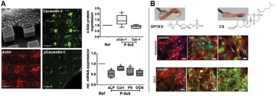
Figure 7.

Biophysical cues from biomaterials for metabolic regulation. A) MG63 cells cultured on micropillars topography with a dimension of 5 µm × 5 µm × 5 µm and a spacing of 5 µm was found to drastically alter actin cytoskeleton organization and to induce attempted caveolae‐mediated phagocytosis of beneath micropillar evidenced by elevated caveolin‐1 expression and activation, which was accompanied with increased ROS and reduced ATP production leading to suppressed osteogenesis, as compared to that on flat surface (Adapted with permission.115 Copyright 2016, Elsevier). B) Supramolecular hydrogels of simple chemical functionality with tunable stiffness were designed to reveal stiffness‐related differentiation of pericytes/MSCs toward different lineages. In combination with metabolomics analysis, two types of lipid, the lysophosphatidic acid (GP18:0) in the glycerolipid pathway and the cholesterol sulfate (CS) in the steroid biosynthesis pathway, were identified and validated as key regulatory metabolites that may be involved in direct chondrogenic (shown as SOX‐9 expression) and osteogenic differentiation (shown as osteopontin expression), respectively (Adapted with permission.116 Copyright 2016, Elsevier).
