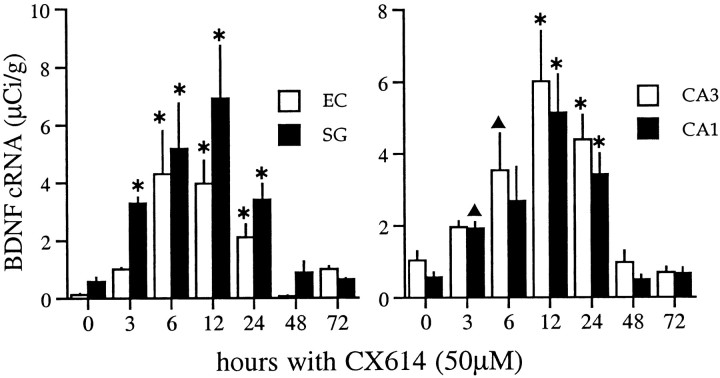
Fig. 5.
Time course of changes in BDNF mRNA content in hippocampal and entorhinal cortex explants treated continuously with 50 μm CX614. Bar graphs show group mean densitometric measures of BDNF cRNA labeling (μCi/gm) within the stratum granulosum (SG) and the superficial entorhinal cortex (EC) (left) and the stratum pyramidale of regions CA3 and CA1 (right). Graphs show the cumulative data from seven experiments (n = 85 explants; group mean values ± SEM) measured from film autoradiograms. Values from vehicle-control slices are presented at the 0 time point; control values represent combined measures from 3, 24, and 72 hr DMSO-treated explants (n = 31). In all fields, there was a significant effect of treatment (p < 0.0001, ANOVA). BDNF cRNA labeling was elevated by 3 hr, was maximal at 12 hr, and was still significantly elevated through 24 hr of treatment. Hybridization densities were not significantly different from control values in slices treated for 48 or 72 hr (*p < 0.001; ▴p < 0.05, for comparison with controls, SNK).

