
Fig. 1.
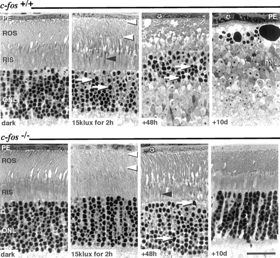
Light microscopy of retinal sections fromc-fos+/+ (top panel) and c-fos−/−(bottom panel) mice: after dark-adaptation (dark), immediately after exposure to 15 klux for 2 hr, and 48 hr and 10 d after light exposure. Retinas of dark-adaptedc-fos+/+ andc-fos−/− mice displayed comparable photoreceptor morphology. Immediately after exposure to 15 klux of white light, ROS showed vesiculation (white arrowheads) in both genotypes, and few RIS appeared condensed (black arrowhead). Forty-eight hours after light exposure, ROS and RIS of c-fos+/+ mice had disintegrated, and the majority of rod nuclei inc-fos+/+ mice showed condensed nuclear chromatin (arrow). Although few condensed RIS (black arrowhead) and darkly stained rod nuclei (arrow) indicated the preceding light exposure, ROS appeared recovered in c-fos−/− mice 48 hr after light exposure. In both genotypes, the presence of phagosomes (*) in the PE indicated the uptake of ROS fragments. Ten days after light exposure, the ONL ofc-fos+/+ mice was amputated, and the INL was in close proximity to the swollen PE. In contrast, photoreceptor and PE morphology inc-fos−/− mice were normal. Stained with methylene blue. Scale bar, 25 μm.