
Fig. 3.
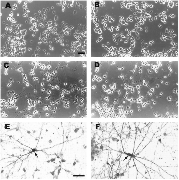
Phase-contrast photomicrographs of treated and untreated, differentiated and undifferentiated PC12 cell cultures (A–D) and of primary cortical neuronal cultures (E, F). A, Undifferentiated, untreated PC12 cell cultures. B, Undifferentiated PC12 cell cultures treated with IL-1β (100 ng/ml for 24 hr). C, Differentiated (NGF-induced), untreated PC12 cell cultures. D, Differentiated PC12 cell cultures treated with IL-1β (100 ng/ml for 24 hr). There are no discernible morphological difference between IL-1β-treated and untreated PC12 cell cultures. E, F, Acetylcholinesterase histochemical reaction of primary cortical neuronal cultures in the absence (E) or presence (F) of IL-1β (100 ng/ml for 24 hr). There was no observable difference in the numbers of AChE-positive cells in response to IL-1β.A–D are the same magnification, and Eand F are the same magnification. Scale bars, 60 μm.