Fig. 3.
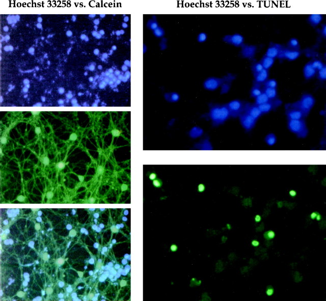
Comparison of methods for assessing apoptosis or cell viability. Left column, Apoptotic CGCs were costained with Hoechst 33258 (top) and 20 μm calcein (middle), which stains only living cells. Superimposition of these images (bottom) demonstrates no colocalization of positive signals, which indicates that Hoechst 33258 does not identify living cells under the conditions used here. Right column, Apoptotic features as assessed by Hoechst 33258 or TUNEL. Hoechst 33258-stained nuclei are condensed as in Figure 2 (top). The same cells labeled by TUNEL show a similar staining pattern (bottom), indicating that both methods provide comparable estimates of apoptosis.

