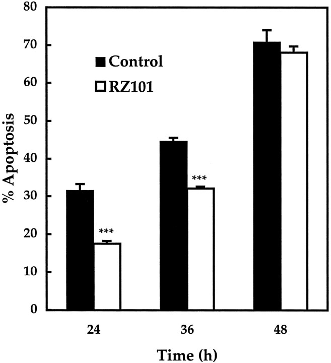
Fig. 8.
Apoptosis in transfected CGCs expressing a ribozyme against rat caspase-3. Apoptosis was assessed after 24, 36, or 48 hr of serum–K+ deprivation. In negative control cells expressing β-galactosidase, apoptosis measured 32 ± 2% (average cells counted, x = 69), 45 ± 1% (x = 89), and 71 ± 3% (x= 38), respectively. In cells expressing RZ101, apoptosis at the same times points was reduced to 18 ± 0.7% (x = 158), 32 ± 0.5% (x = 151), and 68 ± 1.7% (x = 41), respectively. ***p < 0.001 by one-tailed Student'st test (n = 5).

