
Fig. 4.
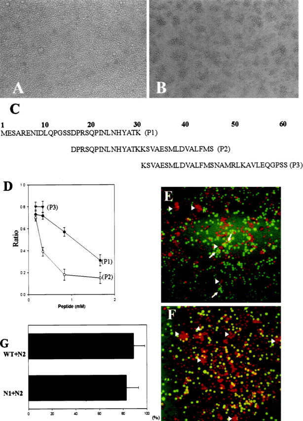
Ninjurin2 mediates homophilic cell adhesion. Native Jurkat cells (A) and Jurkat cells stably transfected with pNINJ2 (B) were resuspended at 1 × 10⁶ cells/ml and allowed to aggregate at 37°C for 1 hr. Note the presence of large aggregates inB. C, List of peptides used in competition experiments and also as antigens to raise anti-ninjurin2 antisera. D, Aggregation assays were performed using ninjurin2 stably expressing Jurkat cells in the presence of each of the indicated peptides. The number of cells in aggregates was determined after 1 hr. The ratio of cells in aggregates to total cells was calculated and plotted versus peptide concentration. Data represents the mean ± SD of three independent experiments. E,F, Aggregation assays were performed with a mixture of N2 cells (stained with red-orange fluorescent dye) and either N1 cells (E) or wild-type cells (F) (stained with greenfluorescent dye). Note that the green cells indicated byarrows and red-orange cells indicated byarrowheads individually form aggregates inE, and only red cells form aggregates inF as indicated by arrowheads.G, Quantitative analysis of aggregation assays using mixed cell populations. The aggregation assays were performed as inE and F, and red cells and green cells in each aggregate were counted. The graph represents the mean ± SD percentage of N2 cells in aggregates that consist predominantly of N2 cells.