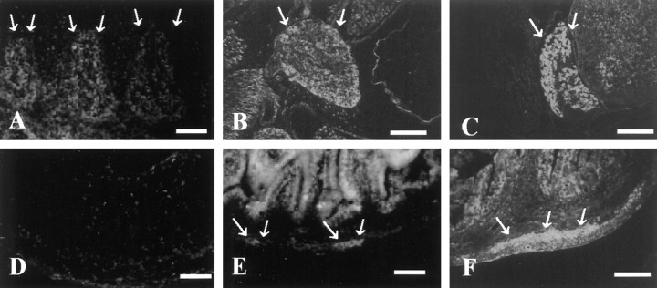
Fig. 7.
Expression of ninjurin2 in sensory and enteric neurons during development. Immunohistochemistry was used to detect ninjurin2 in mouse DRG at E14 (A), E19 (B), and P3 (C) and in mouse enteric plexus at E17 (D), P1 (E), and P3 (F).Arrows denote the ganglia in each micrograph. Scale bars: A, D, E, 50 μm;B, C, F, 150 μm.

