
Fig. 8.
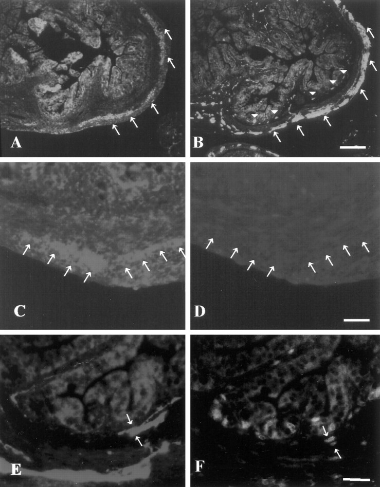
Ninjurin2 is expressed in postmitotic neurons in enteric ganglia. A, B, Ninjurin2 immunoreactivity in enteric plexus in P3 mouse gut (A) was compared with neuron-specific enolase immunoreactivity on an adjacent section (B). Note that myenteric neurons denoted by arrows express ninjurin2, but submucosal neurons denoted by arrowheadsin B, which differentiate later than myenteric neurons, lack ninjurin2 expression in A. Scale bar, 100 μm.C–F, BrdU and either ninjurin2 (C,D) or NSE (E, F) were visualized on the same section of mouse gut at P3. A mouse (P3) was injected with BrdU and killed 1 hr later for immunohistochemistry. Ninjurin2 (C) and NSE expression (E) were visualized by Cy3-conjugated secondary antibody, and proliferating cells were visualized by FITC-conjugated anti-BrdU immunohistochemistry (D,F). The ninjurin2-positive myenteric ganglia lack BrdU staining (arrows in C andD), whereas the NSE-positive cells in the submucosal ganglia indicated by arrows in E and Fare BrdU-positive. Note that C and Drepresent the same section as do E and F. Scale bars, 50 μm.