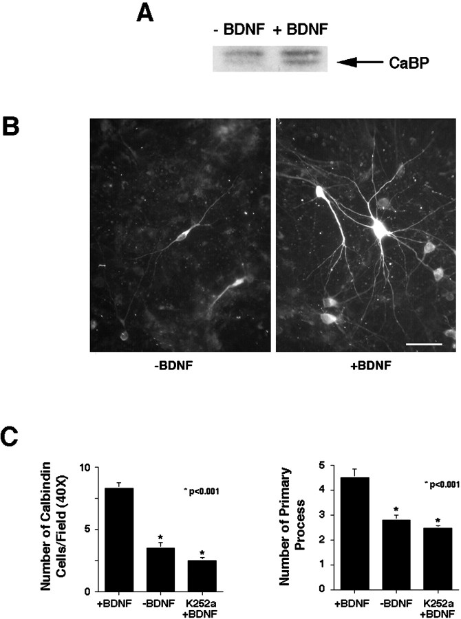
Fig. 5.
The effects of BDNF on calbindin levels in cultured septal neurons. A, Septal neurons from E16 rats cultured for 5 d with 50 ng/ml BDNF produce more calbindin (CaBP) than untreated cells (−BDNF). Fifty micrograms of total protein were loaded in each lane. B, Fluorescent micrograph of calbindin immunoreactivity in cells from septum cultured for 5 d in the presence or absence of BDNF (50 ng/ml).C, Left panel, BDNF treatment led to an increase in the number of calbindin-positive cells in culture, scored per field of view at 40×. Cultures treated with K252a and BDNF showed no increases relative to controls. Right panel, BDNF treatment increased the number of primary neurites emanating from the cell body of calbindin-containing neurons, an effect that was ablated by K252a. Asterisks indicate statistical significance as determined by Student's t tests. Scale bar, 25 μm.

