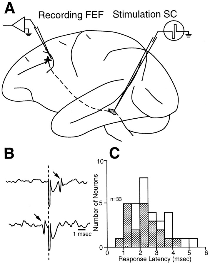
Fig. 1.
Experimental configuration and antidromic responses. A, Lateral view of a rhesus monkey brain illustrating single-neuron recording in the FEF and stimulation of the ipsilateral SC. B, Antidromic response of an FEF neuron (arrow, top trace) and its collision with a spontaneously generated action potential (arrow, bottom trace) that triggered microstimulation. The vertical dashed line indicates the time of SC stimulation.C, Histogram of antidromic latencies for 33 identified corticotectal neurons. Hatched bars indicate saccade-related neurons.

