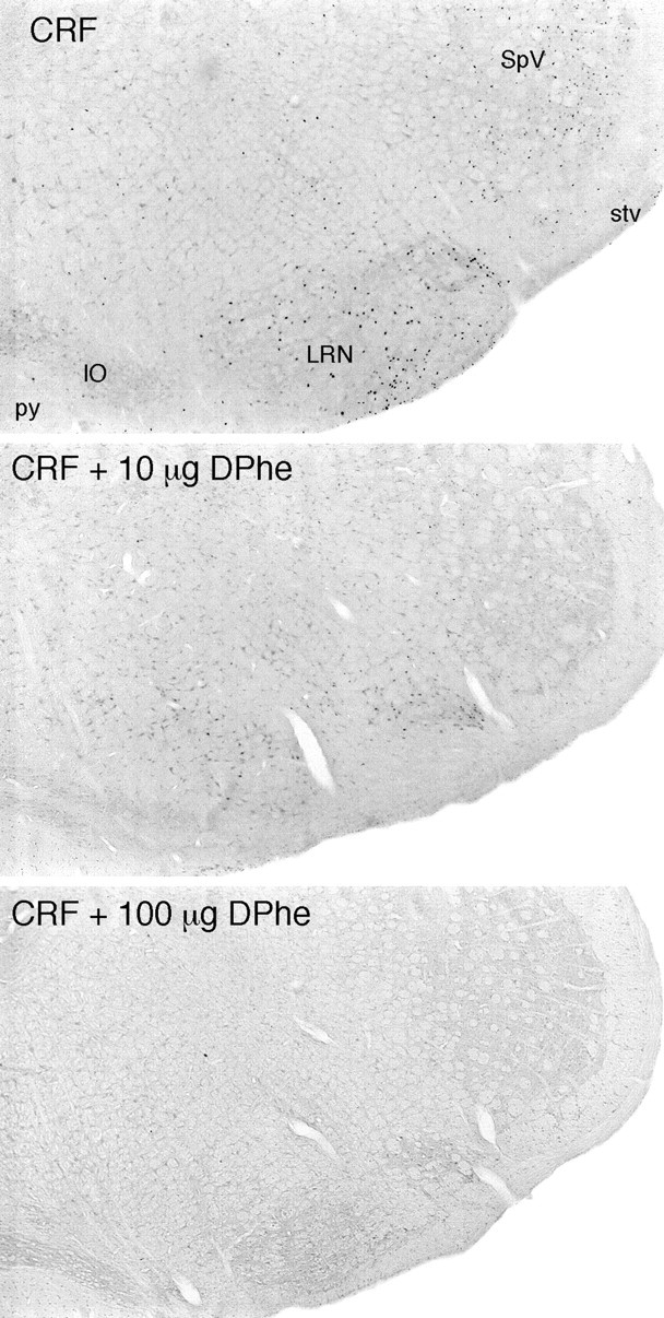
Fig. 2.

Coinjection of a CRF receptor antagonist interferes with central CRF-induced Fos expression in rat brain. Bright-field photomicrographs are of immunoperoxidase preparations to show Fos-ir expression in the ventrolateral medulla of rats that received icv injections of 1 μg CRF alone (top) or with 10 μg (middle) or 100 μg [DPhe12, Nle21,38] r/hCRF12–41. Major sites of peptide-stimulated Fos induction in the lateral reticular (LRN) and spinal trigeminal (SpV) nuclei are markedly diminished in animals coinjected with 10 μg, and essentially abolished in rats treated with 100 μg, of the antagonist. All sections are from animals killed at 2 hr after icv injection, the time of maximal Fos induction in most brain regions. IO, Inferior olivary complex; py, pyramidal tract;stv, spinal tract of the trigeminal nerve. All photomicrographs 30× magnification.
