
Fig. 3.
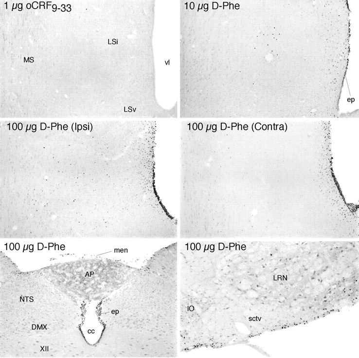
Effects of icv injections at the brain-fluid interfaces. Bright-field photomicrographs show Fos-ir expression in the septal region, near the site of icv injection (top four panels) and caudal brainstem (bottom) in rats killed 2 hr after treatment. Injection of 1 μg oCRF9–33, a peptide fragment that is bound with low affinity by each of the known CRF binding moieties, provokes little evidence of Fos induction even near the site of infusion (top). This contrasts with the effects of 1 μg injections of CRF or UCN (Fig. 7). Injection of 10 μg of the CRF receptor antagonist [D-Phe12, Nle21,38] rat/human CRF12–41(D-Phe) evokes activational responses primarily in the ependymal lining of the ventricular system (ep) near the site of infusion and in immediately adjoining cells but only sporadically in deeper aspects of the brain parenchyma. High doses of the antagonist alone (100 μg D-Phe;middle) produce more robust labeling of the ependyma and periventricular regions, although deep parenchymal labeling is prominent near the site of infusion on the ipsilateral (Ipsi) but not the contralateral (Contra) side of the brain. High doses of the antagonist also result in extensive labeling of the ependyma throughout the ventricular system, as evidenced by labeling seen near the medullary spinal transition area (bottom) and additionally in the meninges (men) and in cells at and just deep to the pial surface of the brain (bottom). Note that labeling at the ependymal and pial surfaces spreads substantially to include cells in deeper regions of the parenchyma only near the site of icv injection.XII, Hypoglossal nucleus; AP, area postrema; cc, central canal; DMX, dorsal motor nucleus; IO, inferior olive;LSv, lateral septal nucleus ventral; Lsi, lateral septal nucleus intermediate; LRN, lateral reticular nucleus; MS, medial septal nucleus;NTS, nucleus of the solitary tract; sctv, spinocerebellar tract; vl, lateral ventricle. All photomicrographs 75× magnification, except bottom right(100×).