
Fig. 5.
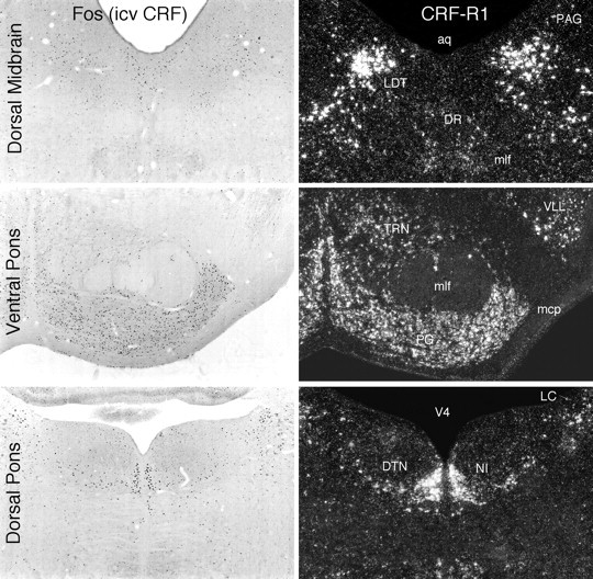
Some brainstem sites of CRF-induced Fos-ir in relation to loci of CRF-R1 mRNA expression. Shown are patterns of Fos induction in brainstem regions seen at 2 hr after icv injection of 1 μg CRF (bright-field, left) and patterns of CRF-R1 mRNA expression in the same regions (dark-field, right). Again, the distributions of the two markers are highly congruent, and most major areas in which Fos induction was detected also express CRF-R1, except for the locus coeruleus (LC), which expresses neither CRF-R, and the dorsal raphé nucleus (DR), aspects of which express CRF-R1 at low levels but CRF-R2 more robustly. aq, Cerebral aqueduct;DTN, dorsal tegmental nucleus; LDT, laterodorsal tegmental nucleus; mcp, middle cerebellar peduncle; mlf, medial longitudinal fasciculus;NI, nucleus incertus; PAG, periaqueductal gray; PG, pontine gray; TRN, tegmental reticular nucleus; V4, fourth ventricle;VLL, ventral nucleus of the lateral lemniscus. All photomicrographs 30× magnification.