Fig. 6.
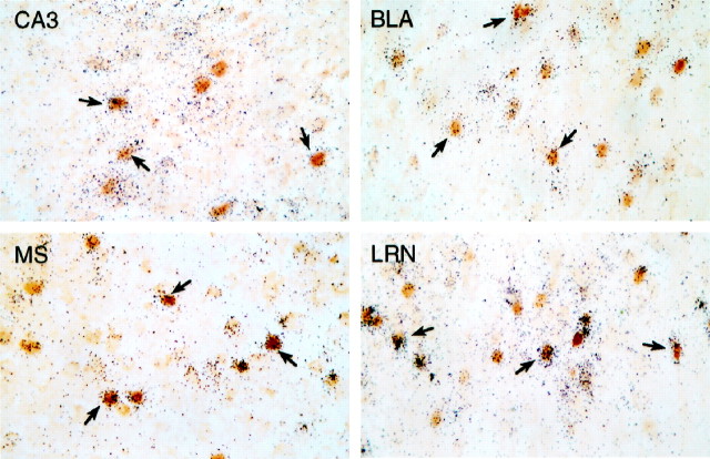
Many neurons that are sensitive to icv CRF injection express CRF-R1 mRNA. Bright-field photomicrographs of combined immunohistochemical and hybridization histochemical preparations show localization of CRF-stimulated Fos-ir (brown nuclei) and CRF-R1 mRNA (blacksilver grains). Overlapping distributions are seen in field CA3 of the hippocampal formation, basolateral amygdaloid (BLA), medial septal (MS), and lateral reticular (LRN) nuclei, among many other regions. Examples of doubly labeled cells are indicated (arrows). All photomicrographs 300× magnification.

