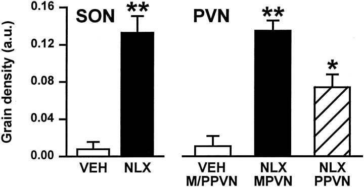
Fig. 1.
Changes in expression of c-fos mRNA in the supraoptic and paraventricular nuclei during morphine withdrawal. Mean silver grain density [arbitrary units (a.u.) + SEM] in c-fos-labeled sections through the SON and PVN (magnocellular and parvocellular divisions, MPVN and PPVN, respectively) of morphine-dependent rats 45 min after vehicle (VEH; 0.15 m saline, s.c.; n = 4, magnocellular and parvocellular paraventricular nuclei combined) or naloxone (NLX; 5 mg/kg, s.c.; n = 4) administration. *p < 0.05, **p < 0.01 versus vehicle; Student'st test.

