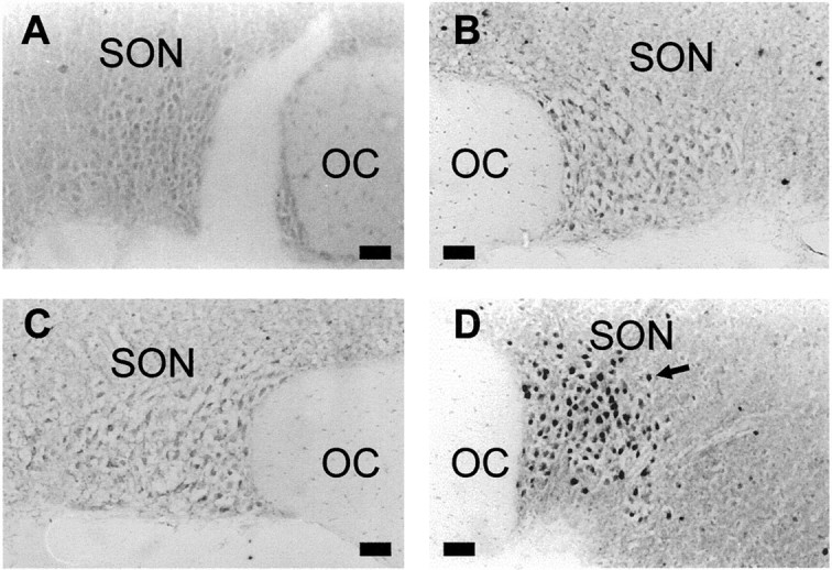
Fig. 2.

Fos protein in the supraoptic nuclei of morphine-naïve and -dependent rats. Photomicrographs of coronal sections through the SON processed for Fos immunohistochemistry from a morphine-naïve rat given vehicle (0.9% saline, 0.5 ml/kg, s.c.) (A), a morphine-naïve rat given naloxone (5 mg/kg, s.c.) (B), a morphine-dependent rat given vehicle (C), and a morphine-dependent rat given naloxone (D). Nuclei immunoreactive for Fos protein appear as intense black staining (arrow). OC, Optic chiasma. Scale bars, 50 μm.
