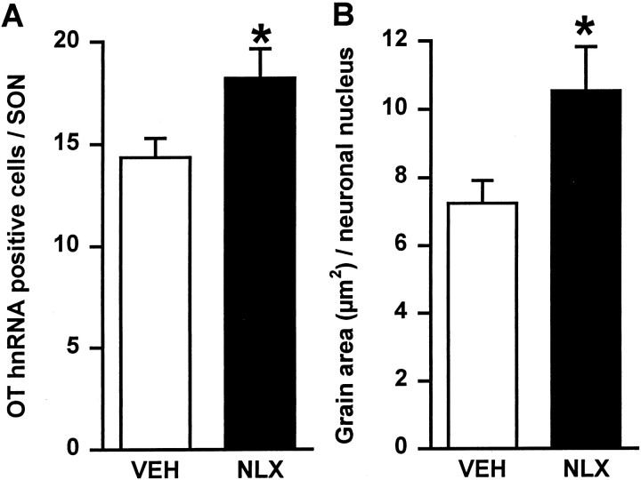
Fig. 5.
Changes in expression of oxytocin hnRNA in supraoptic nucleus cells during morphine withdrawal. Coronal sections containing the supraoptic nucleus from rats treated with either chronic morphine and acute subcutaneous 0.15 m saline (VEH; n = 5) or chronic morphine and acute naloxone (NLX; 5 mg/kg, s.c; withdrawn,n = 6) were hybridized with a3H-labeled cDNA probe complementary to 210 bases of intron 1 of rat oxytocin hnRNA. A, Mean + SEM number of oxytocin hnRNA-positive cells per supraoptic nucleus section.B, Mean + SEM silver grain area per supraoptic nucleus neuronal nucleus. *p < 0.05; Student'st test.

