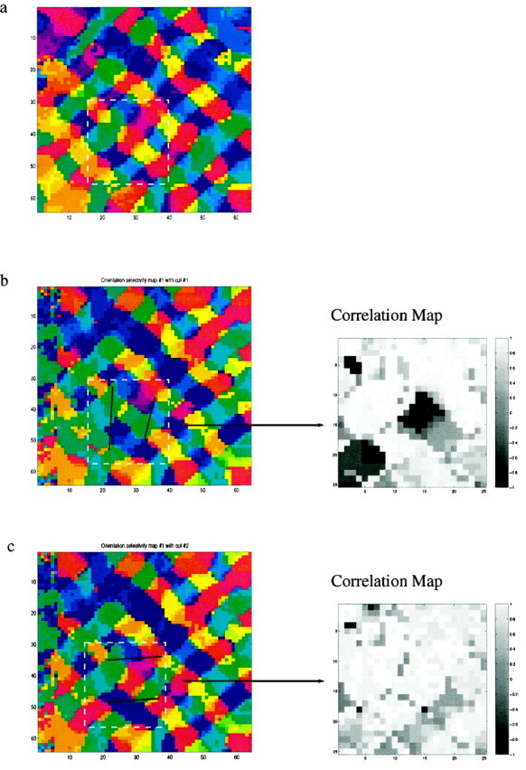
Fig. 6.

Proposed experimental test for the scaffold hypothesis. We expect that the location and orientation of small cuts would affect the layout of the orientation map that develops during the reverse suture phase. a, Trained map after MD, as in Figures 2b and 4a. Before RS, selective small cuts are made to the cortical surface, disrupting lateral connections. After RS, the maps are imaged. b, Black lines indicate two cuts designed to sever many of the horizontal lateral connections to a region preferring horizontal orientations (red). The cuts created a significant difference in the preferred orientation after RS. On the right a circular correlation map, in a region of interest, between the uncut map (Fig. 5a) and the cut map is shown. Small values are indicative of significantly different preferred orientations. c, Control experiment, in which two cuts do not disrupt the horizontal connections. In this case, we anticipate a much smaller change in the orientation map, indicated by the absence of small values in the correlation map.
