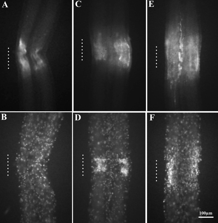
Fig. 1.
Appearance of eNOS immunoreactivity and microglial cell accumulation at sites of CNS damage. The connectives connecting ganglia are paired, with the bundled axons of each connective surrounded by a cellular sheath. Adult leech nerve cords were crushed and double-stained with a monoclonal antibody against human eNOS and with the fluorescent nuclear dye Hoechst 33342. The approximate longitudinal extent of the crushes is indicated by vertical dotted lines. Five minutes after crushing, eNOS immunoreactivity was present among nerve fibers in the connectives (A), but microglial cells were still evenly distributed throughout the connectives (B), resembling controls. Note that immunoreactivity was predominantly where crushed axons, glia, and microglia were located and not in the sheath. Three hours after injury, eNOS immunoreactivity persisted at the lesion (C), and microglial cells had accumulated (D). After 24 hr, eNOS immunoreactivity was more diffuse and expanded outside the crush (E). Microglial cells also occupied a larger area (F).

