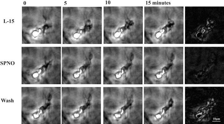
Fig. 8.
NO donor SPNO reversibly arrested movement of microglial cells in three-dimensional collagen gels. Microglia that migrated into the gels moved their lamellipodia with little translocation. Phase contrast images in each row were separated by 5 min intervals; right panels are computer-generated subtractions of the last two images and show cell movement in 5 min in white. Addition of SPNO to a final concentration of 1 mm in L-15 culture medium arrested movement of lamellipodia (middle row), whereas addition of aged NO donor solutions (data not shown) was without effect. The usual motility resumed within minutes of washout (bottom row).

