Fig. 3.
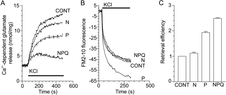
Activation of VDCCs coupled to exocytosis also inhibits endocytosis. A, Ca2+-dependent glutamate release evoked by 30 mm KCl in the presence of 1 μmω-conotoxin-GVIA (N), 30 nmω-agatoxin-IVA (P), or 5 μmω-conotoxin-MVIIC (NPQ); n = 4 (±SEM). B, Nerve terminals were loaded for 2 min with FM2–10 during S1 stimulation using KCl (30 mm) in the presence of N-, P-, or N/P/Q-type VDCC blockers. A representativetrace of the subsequent Ca2+-dependent release of loaded FM2–10 (S2) by a standard stimulus of 30 mm KCl is displayed. Exocytosis is constant during S2 stimulation; therefore, endocytosis is calculated as Ca2+-dependent fluorescence decrease in the S2 stimulation; n = 3–4. In all experiments, VDCC antagonists were present 2 min before stimulation. KCl stimulation is represented by the bar. C, KCl-stimulated retrieval efficiency in the presence of VDCC antagonists normalized to control [n = 4 (±SEM)].

