
Fig. 3.
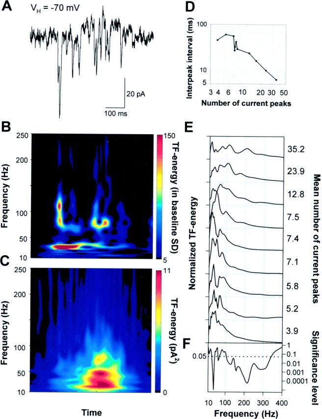
During spontaneous network events, AMPA–kainate receptor-mediated currents show oscillations at gamma- and high-frequencies. A, A spontaneous event in a P5 pyramidal neuron clamped at −70 mV showing the AMPA–kainate receptor-mediated glutamatergic currents. B, A TF representation of the glutamatergic currents in A. Time runs along the x-axis and frequency along they-axis. The time scale is the same as in the current trace above the TF representation. The amplitude is coded in color so that red color corresponds to higher amplitude (see Materials and Methods for details). The high-frequency components are related to the current clusters occurring at gamma frequencies.C, An averaged TF representation of 103 consecutive spontaneous bursts of AMPA–kainate currents displaying the major peak in the lower (20–40 Hz) and a subsidiary peak in the upper (50–80 Hz) gamma-frequency band. D, The mean interpeak interval was dependent on the mean number of AMPA–kainate current peaks in bursts (p < 0.005; one-way ANOVA).E, Frequency distributions of the AMPA–kainate receptor-mediated current bursts in nine neurons. The distributions were obtained by averaging the TF representations of all current bursts (33–103 averages) in a single recording and then averaging 25–100 msec around the major peak. The distributions are normalized to have unit area. The mean number of current peaks during a population burst is given right to the distributions. F, Significance level as a function of frequency (one-way ANOVA). A below average amount of AMPA–kainate inputs per burst was associated with pronounced gamma-frequency oscillations, whereas large numbers of AMPA–kainate inputs/burst were found in neurons showing prominent high-frequency oscillations.