Fig. 2.
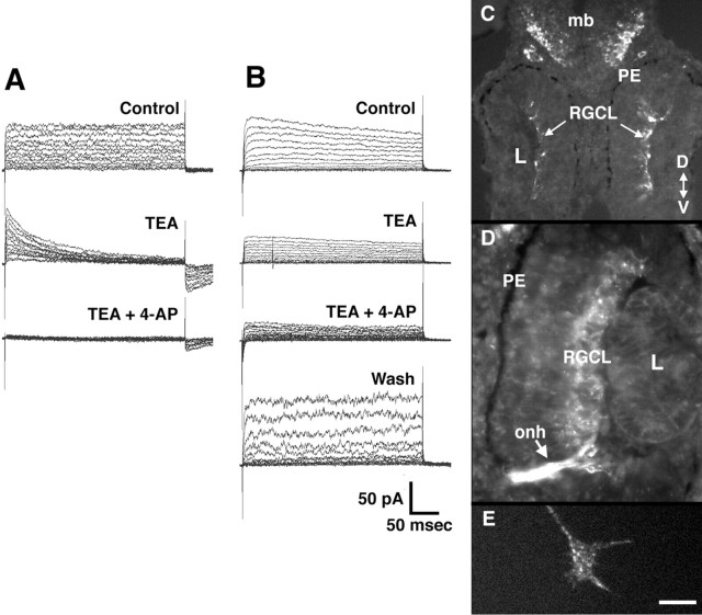
Developing RGCs express Kv channels. A, B, Kv currents recorded from two different stage 33/34 equivalent RGCs in culture in the whole-cell configuration (see Materials and Methods). The cells were held at holding potential of −80 mV, and 400 msec voltage steps were applied in 10 mV increments from −60 to +70 mV. In both cells, outward Kv currents are observed that are sensitive to both 3 mm 4-AP and 50 mmTEA. With the cell shown in B, wash out with control solution was able to reverse the blockade. C–E,Immunolabeling with a rabbit polyclonal antibody against rat Kv4.3.C, D, Transverse sections through stage 33/34 (C) and stage 37/38 (D) retinas showing labeling of cells in the RGC layer. PE, Pigment epithelium; L, lens; onh, optic nerve head; mb, midbrain; RGCL, RGC layer; D, dorsal; V, ventral.E, An RGC growth cone in culture immunolabeled with the Kv4.3 antibody. The body of the growth cone, the filopodia, and the lamellopodia are labeled in a punctate fashion. Scale bar (shown inE): C, 50 μm;D, 25 μm;E, 5 μm.

