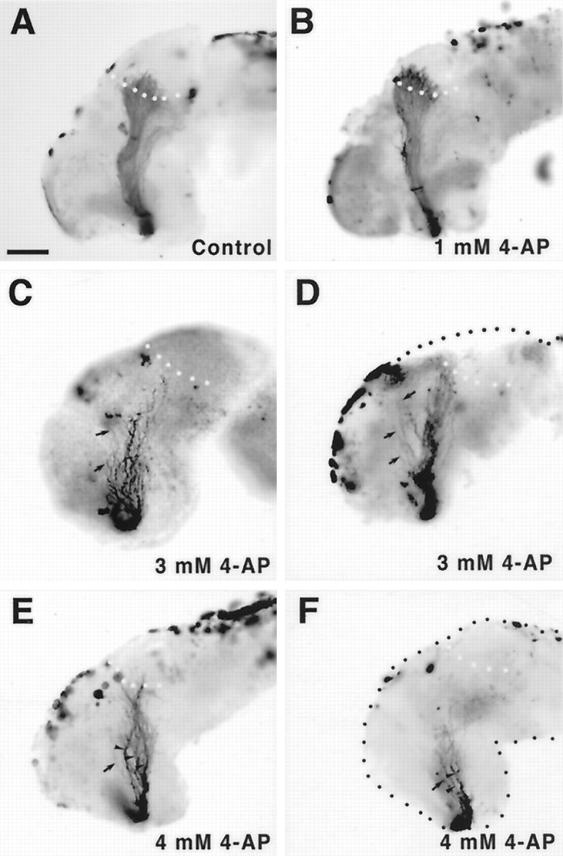
Fig. 3.

4-AP disrupts the optic projection.A–F, Whole-mount brain preparations showing HRP-labeled optic projections in control (A) and 4-AP-treated (B–F) brains. At a low concentration of 4-AP of 1 mm (B), optic axons behave normally. Optic projections exposed to higher levels of 4-AP, 3 mm (C, D) and 4 mm (E, F), are shorter than control, appear defasciculated (arrowheads), and have many axons that grow aberrantly away from the optic tract (arrows). Scale bar (shown inA), 100 μm.
