
Fig. 2.
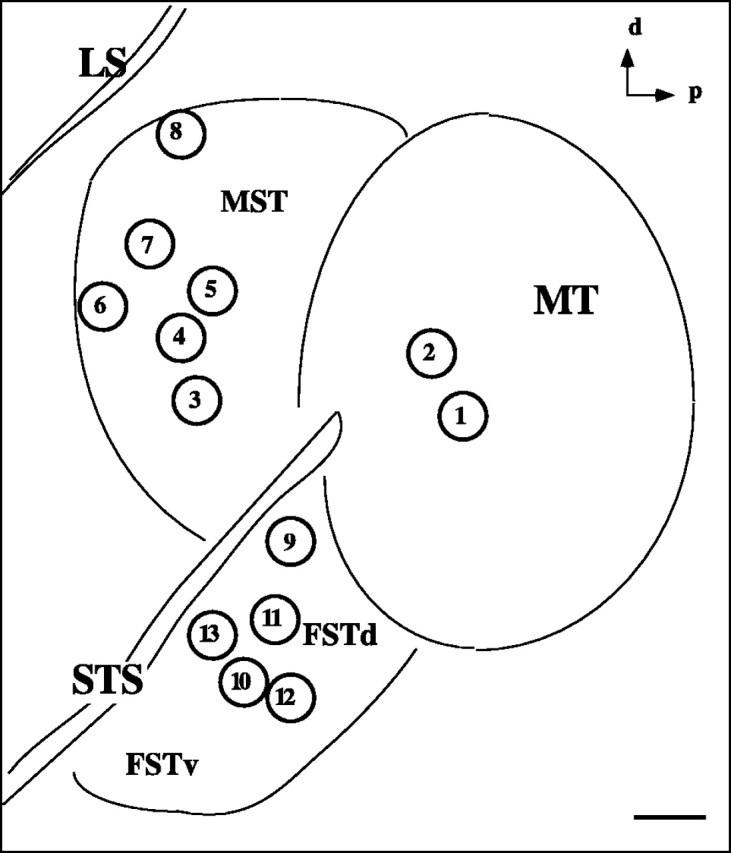
Summary of injection sites. Eachcircle represents the approximate location of an injection made in this study. Sites 1 and2, within MT, were injections of anterograde tracer; the others were injections of different retrograde tracers into MST and FST. See Table 1 for details on the nature of the injections and the results. d, Dorsal; p, posterior;LS, lateral sulcus; STS, superior temporal sulcus; MST, medial superior temporal area;FSTd,FSTv, dorsal and ventral subdivisions, respectively, of the fundus of the superior temporal sulcus. Scale bar, 1 mm.