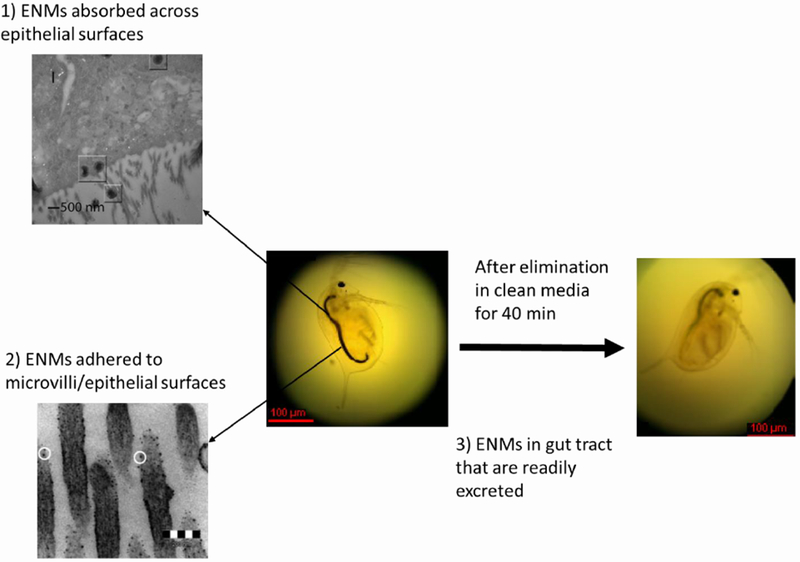
Figure 4:

Fractions of engineered nanomaterials (ENMs) that can be detected in organisms with a digestive tract: 1) ENMs absorbed across epithelial surfaces; this figure (upper left) shows carbon nanotubes (CNTs) that had been absorbed by microvilli (see squares) although additional analysis using high resolution transmission electron microscopy (HRTEM) revealed that these particles were amorphous carbon and not CNTs.11 2) ENMs adhered to microvilli; this figure (bottom left) shows apparent fullerene particles adhered to the microvilli.12 3) ENMs in gut tract that are readily excreted; this figure (far right) shows that the gut tract of the Daphnia magna turned from black (as a result of uptake of few layer graphene for 24 h) to transparent or green after an elimination period of 40 min with algae feeding;256 adapted with permission from 256 2013 American Chemical Society.
