
Fig. 7.
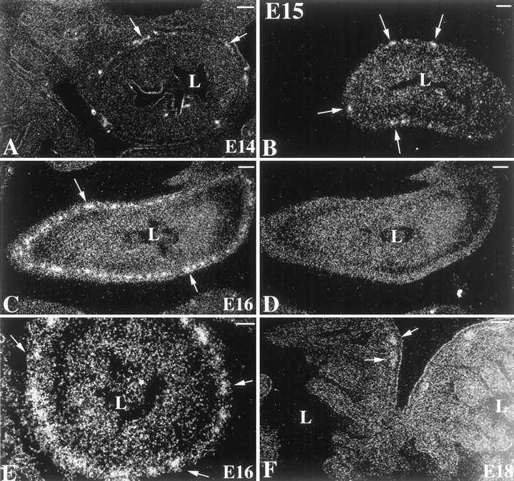
Expression of the 5-HT2B receptor is developmentally regulated in the ENS. mRNA encoding the 5-HT2B receptor was located in fetal tissue by in situ hybridization with an antisense 35S-riboprobe. As a control, alternate serial sections were hybridized with a sense35S-riboprobe. Sections are visualized by using reflected dark-field illumination. A, E14 fetal bowel (antisense riboprobe). mRNA encoding the 5-HT2B receptor is found in scattered ganglia of the primordial myenteric plexus in the outer gut mesenchyme (→). The dark-field bright material lining the lumen (L) is caused by the chemographic effects of meconium and is not specific labeling. The section was exposed for 12 weeks. B, E15 (antisense riboprobe). The degree of labeling is approximately equal to that seen at E14; however, exposure for only 8 weeks is required to detect mRNA encoding the 5-HT2B receptor in primordial myenteric ganglia (→).C, E16 small intestine (antisense riboprobe). mRNA encoding the 5-HT2B receptor is detectable in many myenteric ganglia (→) that surround the gut. D, E16 control (sense riboprobe). An adjacent section, serial to that illustrated in C, is shown. No structures are labeled.E, E16 colon (antisense riboprobe). Labeled ganglia (→) are even more numerous than in the small intestine at the same age. F, E18. Only occasional ganglia are labeled (→). Scale bars, 10 μm.