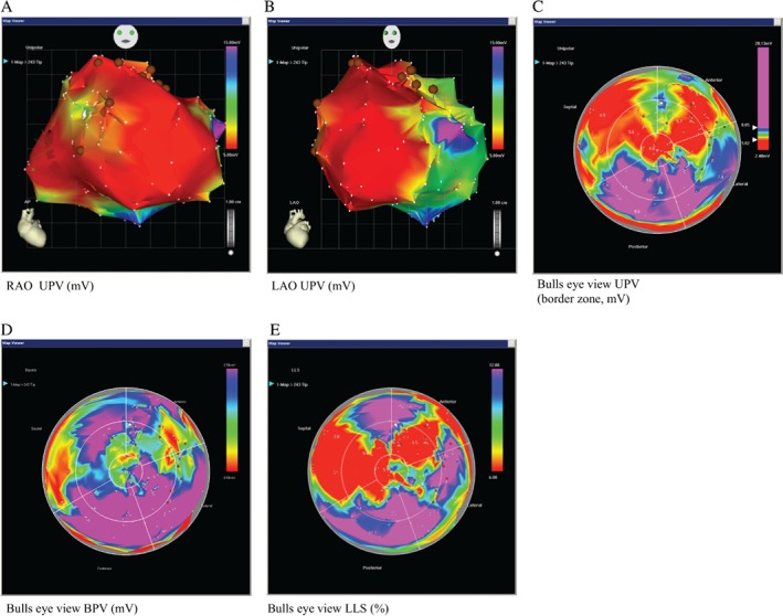
Figure 2.

Exemplary images of the NOGA procedure of a 69‐year‐old man with ischaemic heart failure. Three‐dimensional left ventricular mapping performed using the NOGA XP® system. (A) Unipolar voltage (UPV) in right anterior oblique (RAO) (UPV 5–15 mV), (B) left anterior oblique (LAO) (UPV 5–15 mV) and (C) in bulls eye projections (UPV with focus on the border zone 5–8 mV) are displayed with margins manually set to standardized values. (D) Scar definition using bipolar voltage (BPV 0.5–2.5 mV) and (E) local linear shortening (LLS, 6–12%). Colour encoding is displayed at the right upper border of the pictures. White points indicate mapping points and brown dots indicate locations for injections in the infarction border zone. A mismatch between UPV, BPV and LLS is observed showing maintained electrical viability (UPV, BPV) in certain areas of the ventricle with no systolic movement (LLS), suggestive of hibernating myocardium; when scar is not transmural, a mismatch between UPV and BPV is seen, two injections were placed in this specific area; compare posterior area of map in the bulls eye perspective (viable and contractive) to the anterior‐septal region.
