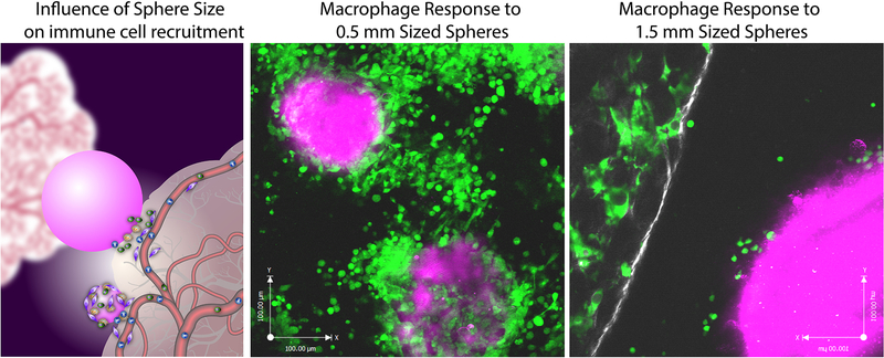
Figure 4.
Extravasation of macrophage cells from peripheral tissue on to spherical materials of 0.5 mm and 1.5 mm spheres. (a) schematic describing how smaller spheres better conform to peripheral adjacent tissue cervices compared to larger spheres and this influence on the ability of immune cells to extravasate on to implants from vasculature. In vivo intravital imaging of macrophage behavior and accumulation at 7 days post-implantation on to (b) 0.5 mm and (c) 1.5 mm diameter sized Ba+ crosslinked alginate spheres. (macrophages depicted in green, peripheral tissue in white and implanted spheres in magenta). Figure and caption adapted with permission from reference [25].

