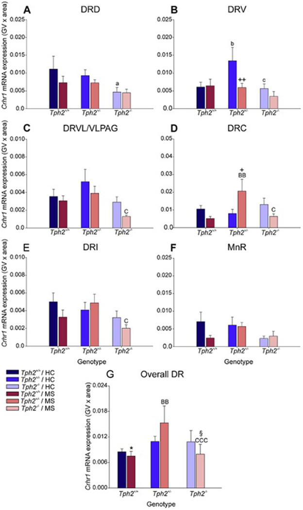
Figure 7.

Effects of maternal separation in wild-type and Tph2-deficient mice on overall Crhr1 mRNA expression within each subregion of the dorsal raphe nucleus (DR) and median raphe nucleus (MnR). Graphs represent the mean ± SEM of Crhr1 mRNA expression in the (A) dorsal raphe nucleus, dorsal part (DRD), (B) dorsal raphe nucleus, ventral part (DRV), (C) dorsal raphe nucleus, ventrolateral part (DRVL), (D) dorsal raphe nucleus, caudal part (DRC), (E) dorsal raphe nucleus, interfascicular part (DRI), (F) MnR, and (G) across all subregions in the DR and MnR. Male wild-type, heterozygous, and homozygous Tph2 knockout mice were exposed to either animal facility-reared control conditions (HC) or maternal separation (MS), consisting of daily separation (3 h) of mixed litters from the dam postnatal day 2 (P2) through P15, inclusive. Sample sizes per treatment group: Tph2+/+ HC (n = 8); Tph2 HC (n = 8); Tph2−/− HC (n = 6); Tph2+/+ MS (n = 7); Tph2+/− MS (n = 8); Tph2−/− MS (n = 6); (N = 43). Post hoc comparisons were made using Fisher’s least significant difference (LSD) tests; *p < 0.05, Tph2+/+ MS versus Tph2+/+ HC; +p < 0.05, ++p < 0.01, Tph2 MS versus Tph2 HC; §p < 0.05, Tph2−/− MS versus Tph2−/− HC; ap < 0.05, Tph2+/+ HC versus Tph2−/− HC; bp < 0.05, Tph2+/+ HC versus Tph2+/− HC; cp < 0.05, Tph2+/− HC versus Tph2−/− HC; BBp < 0.01, Tph2+/+ MS versus Tph2−/− MS; Cp < 0.05, Tph2+/− MS versus Tph2−/− MS.
