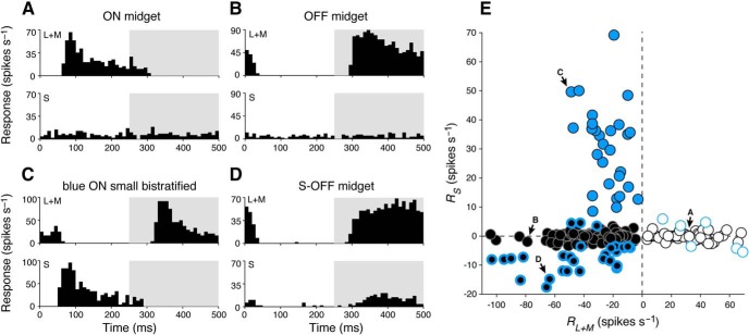
Figure 5.
Cone-mechanism-specific responses in retinal ganglion-cell subtypes. A, Peristimulus time histogram of a typical ON midget ganglion cell shows spiking activity in the ON phase (0–250 ms) of a 2 Hz modulating (L+M)-cone-isolating stimulus (top) while showing no activity during a modulating S-cone-isolating stimulus (bottom). B, A typical OFF midget cell shows the reverse behavior, with spiking activity in the OFF phase (250–500 ms) of a 2 Hz modulating L+M cone-isolating stimulus (top) but also showing no activity during a modulating S-cone-isolating stimulus (bottom). C, A blue ON ganglion cell responds to both L+M (top) and S (bottom) cone-isolating stimuli, with opposite phase. D, An S-OFF midget ganglion cell demonstrates classical sensitivity to L+M-cone stimuli (top), as well as a small response to S-cone stimuli (bottom); both responses occur in the OFF phase. E, Ganglion-cell types cluster based on their responsiveness to S-cone (RS) and L+M-cone (RL+M) stimulation. Blue circles, blue ON cells; white circles, ON midget cells; black circles, OFF midget cells. Blue-bordered white and black circles denote midget ganglion cells with |RS| > 3.5 spikes/s. Labeled cells are those shown in A–D, respectively.

