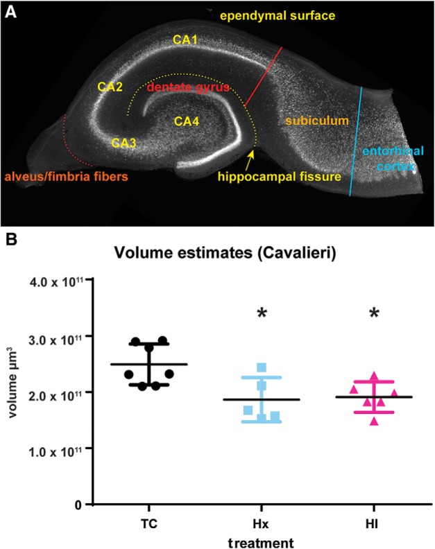
Figure 1.
Volumetric changes were observed in the hippocampal and dentate gyri in response to HI calculated by the Cavalieri estimation. A, NeuN staining of a transverse slice through the hippocampal formation reveals the neuronal subfields of the fetal sheep hippocampal and dentate gyri in the stereotypical “jelly-roll” configuration. Areal calculations of the CA1-CA4 subfields and the dentate gyrus of each slice excluded the subiculum and the entorhinal cortex at a border drawn tangential to the end of the CA1 neuronal subfield (red solid line) between the ependymal surface of the hippocampal gyrus and the hippocampal fissure (yellow dotted line), and the alveus was also excluded where it coalesces to form the fimbria fibers (orange dotted line). B, After 4 weeks, the hippocampal volume of fetuses exposed to Hx or HI was reduced versus controls (TC). No significant differences were observed between Hx and HI treatments. *p < 0.05 (Tukey's post hoc multiple-comparison test).

