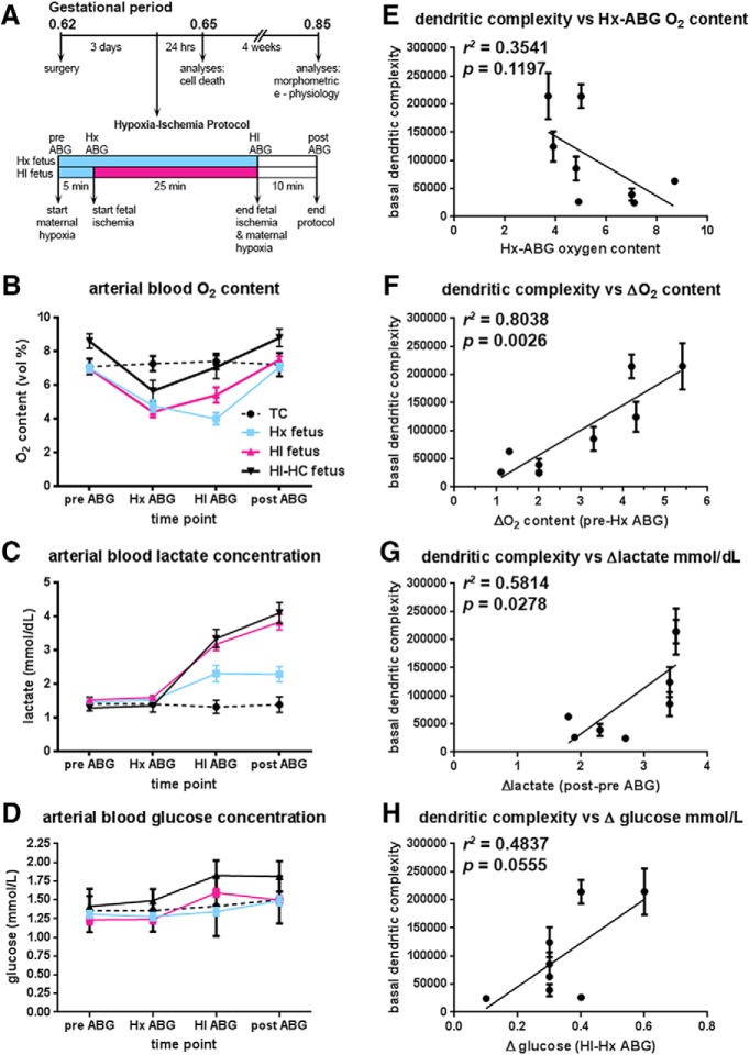
Figure 4.
Fetal ABG values for treatment groups during experimental paradigm. A, Flow chart of the experimental protocol described in Materials and Methods. B, Fetal oxygen content (vol %) initially drops in response to maternal hypoxia. With the onset of fetal ischemia, fetal oxygen content in both HI groups (red and solid black lines) attempts to rebound back to baseline levels, while remaining relatively constant in the fetuses exposed only to maternal hypoxia (Hx group blue line). Within 10 min of the cessation of maternal hypoxia and fetal ischemia, fetal oxygen content returns to baseline levels. The differences in oxygen content between the HI and HI-HC groups reflect the sampling site from which the blood gas was collected; in the fetus, oxygen content is higher in carotid arteries than in femoral arteries. Black dotted lines indicate normal control fetuses (TC). C, Fetal lactate concentration increases markedly in HI groups (red and solid black lines) by the end of the ischemic period and continues to rise 10 min after the cessation of fetal ischemia and maternal hypoxia compared with fetuses exposed only to maternal hypoxia (Hx group, blue lines). D, Fetal glucose concentration rises markedly in HI groups (red and solid black lines) compared with Hx group (blue line). E, The basal dendritic complexity of HI-treated CA1 hippocampal neurons is plotted versus the fetal systemic oxygen content 5 min into maternal hypoxia. F, Basal dendritic complexity of HI-treated CA1 hippocampal neurons is positively associated with the magnitude of the initial fetal response to maternal hypoxia defined as the difference between the pre-ABG (baseline) and Hx-ABG oxygen content values. The greater the drop in the oxygen content value from baseline for the HI-HC fetus, basal dendritic complexity is observed to increase in CA1 neurons. G, Basal dendritic complexity of HI-treated CA1 hippocampal neurons is positively associated with the magnitude of change in lactate recovery defined as the difference between post-ABG (recovery) lactate value and the pre-ABG (baseline) lactate value. H, Basal dendritic complexity of HI-treated CA1 hippocampal neurons versus the magnitude of change in fetal systemic glucose, defined as the difference between the HI-ABG glucose value and the Hx-ABG glucose value.

