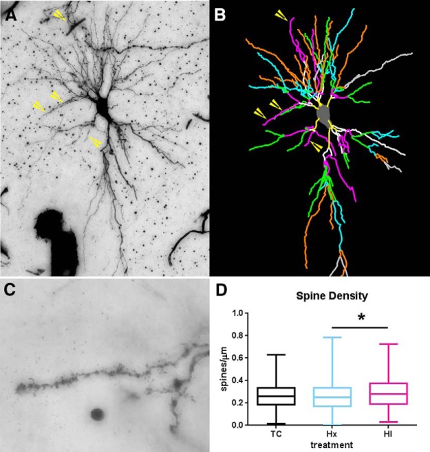
Figure 6.
Augmented basal dendritic arbor maturation is accompanied by no changes in basal dendritic spine density. For the same population of CA1 neurons that were sampled for dendritic morphology, spine density was quantified on third-order terminal dendritic branches of all experimental groups of neurons. A, B, Example of third-order terminal branches (yellow arrows) that were present on the Golgi-impregnated neuron (A) and the corresponding Neurolucida tracing (B, purple/hot pink lines). B, Branch order rank is indicated by color: bright yellow represents first order; white represents second order; purple/hot pink represents third order; bright green represents fourth order; cyan blue represents fifth order; orange represents sixth order; slate gray represents seventh order. C, Dendritic spines visualized on a Golgi-impregnated CA1 hippocampal pyramidal neuron. D, Spine density in control, hypoxia-only (Hx), and HI groups at 4 weeks after treatment. Preterm neurons in both the Hx and HI groups (blue and red bars) revealed no significant change in the number of spines versus controls (black bars). However, Hx- versus HI-treated groups were significant at p < 0.05 (Dunn's correction for multiple pairs, black horizontal bar with asterisk). n = 354 TC, 343 Hx, and 341 HI tertiary basal dendrites. *p < 0.05.

