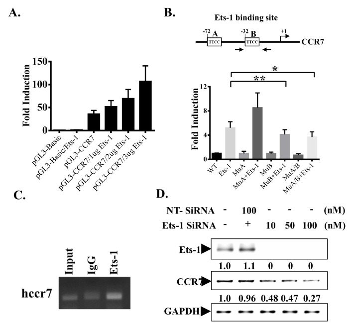
Fig. 2.
Ets-1 binds to CCR7 promoter and controls its expression. (A) MDA-MB-231 cells were transfected with indicated plasmids. Cells were harvested after 24 hours and the promoter activity was examined by luciferase assay. (B) 2 Ets-1 binding sites on the CCR7 promoter region are shown. These sites were either mutated separately (MuA or MuB) or together (both mutated, MuA/B). The promoter activity was examined by luciferase assay and shown in the lower panel. (C) MDA-MD-231 cells were subjected to ChIP analysis. Chromatin was precipitated with an anti-Ets-1 antibody or an IgG antibody as a negative control. Precipitated DNA was amplified using primers (Fig. 2B, arrows) specific for a 220 bp fragment of the CCR7 promoter spanning the −32 Ets-1 binding sites. As a positive control, total DNA was diluted 1:100 and subjected to PCR (input). (D) MDA-MB-231 cells were transfected with indicated amounts of Ets-1 siRNA and harvested after 24 hours. Whole-cell lysates were prepared and subjected to Western blot analyses with indicated antibodies.

