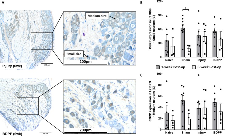
Fig 7. IVD injury and dietary polyphenols of BDPP did not affect the CGRP expression in DRG.
(A) Representative L2 DRGs (collected at 6 weeks post-op) with CGRP-immunopositive neurons (stained brown color) from Injury and BDPP groups. Small- and medium-sized neurons, which corresponds to unmyelinated c-fiber and myelinated a-delta fiber, respectively, were determined. (B) and (C) Percentage immunopositivity of CGRP from small- and medium-sized neurons, respectively, at 1-week (n = 6 for Naïve, n = 8 for Sham, n = 7 for Injury & n = 7 for BDPP) and 6-week post-op (n = 3 for Naïve, n = 3 for Sham, n = 6 for Injury & n = 4 for BDPP). Scatter plots present the CGRP immunopositive neurons from individual animals. Cross bars present group mean±SEM values. * indicates P<0.05 between groups.

