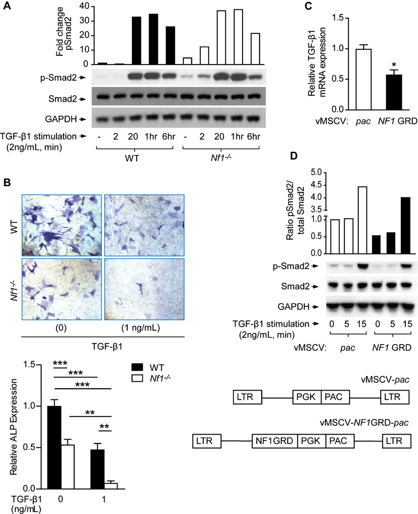
Fig. 2.
Nf1-deficient MSCs exhibit hyperactivation of the Smad pathway and impaired osteoblast differentiation in response to TGF-β1. (A) p-Smad2, Smad2, and GAPDH levels were detected by Western blot in MSCs stimulated with TGF-β1. The quantitative fold change in p-Smad2 was determined relative to the loading control, as shown in the bar graph. The experiment was repeated on three independent occasions with similar results. (B) MSCs were cultured in osteogenic differentiation medium supplemented with TGF-β1. Representative photomicrographs (top panel) show alkaline phosphatase (ALP)-positive osteoblasts (magnification × 200). ALP expression was quantified and normalized to the WT control as shown (bottom panel). n = 3 technical replicates. The assay was repeated twice with similar results using independent cell lines. **p < 0.01, ***p < 0.001. (C) TGF-β1 mRNA expression was measured in Nf1−/− MSCs following retroviral transduction with control vector (MSCV-pac) versus the functional, full-length NF1 GRD construct. n = 3 technical replicates. The experiment was repeated on three independent occasions with similar results. *p < 0.05. (D) p-Smad2, Smad2, and GAPDH levels were detected by Western blot in MSCs stimulated with TGF-β1 following transduction with either MSCV-pac or MSCV-NF1 GRD retroviral vectors. The quantitative fold change in p-Smad2 was determined relative to the level of total Smad2 protein, as shown in the bar graph.

