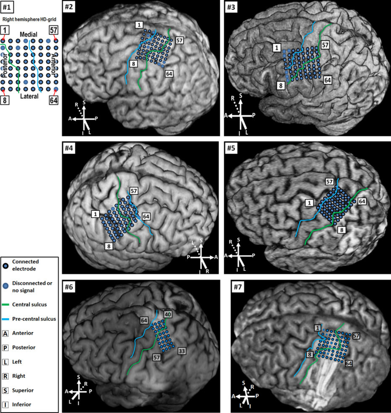
Fig. 3.
Location of HD-ECoG grids. For Subjects 2–5, locations were determined by the MRI-CT co-registration approach (see Section 2.4.1). For Subjects 6 and 7, grids could be visualized on MRI alone, thus co-registration with CT was not necessary. For Subject 1, MRI scan was not available due to the presence of metal inside his body. Thus, localization was performed by identifying the central sulcus location using pre- and post-implantation CT scans, and the electrode locations relative to the central sulcus and other anatomical landmarks are portrayed here.

