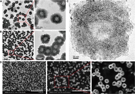
Fig. 2. Morphology characterization of the self-assembled nanostructures (nanosunflowers).

(A) TEM (200 kV) images of the nanosunflowers with enlarged structural details. (B) Bio-TEM (80 kV) images with enlarged polymer structural details. (C) High-resolution TEM (200 kV) images showing the distribution of ultrasmall NPs on the self-assembled nanostructure. (D) SEM images with enlarged surface topography of the nanosunflowers.
