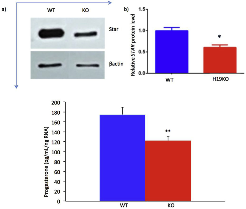
Fig. 1. Decreased STAR expression and progesterone in primary granulosa cells of H19KO mice.
a) 5 week old H19KO and WT C57/Bl6 female mice (n = 3 per group) were superovulated by intraperitoneal injection of 5IU pregnant mare serum gonadotropin (PMSG) × 2 to stimulate follicle development. 24 h after the second injection, mice were given an injection of 5IU hCG, and GCs were isolated 16 h later and cultured in α-MEM. After 24 h of culture, Western blot was performed to measure STAR protein levels, using β-actin as control. Decreased STAR was observed in primary GCs from H19KO mice as compared to WT. b). Quantification of Western blot showing decreased STAR expression; p < 0.05. c) Progesterone concentration was determined via ELISA after 24 h of culture and normalized to total RNA. Progesterone production from cultured H19KO GCs was decreased as compared to WT GCs. p < 0.01. All data were analyzed using Student’s t-test. *p < 0.05; **p < 0.01.

