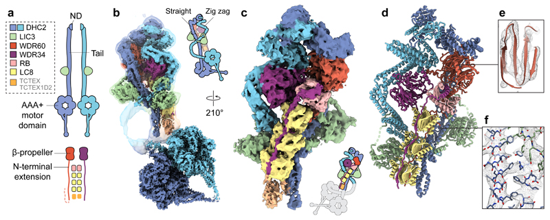
Figure 1. Cryo-EM structure of the dynein-2 complex.
(a) Overview of the human dynein-2 subunits and their stoichiometry in the structure, coloured according to the code in the upper left. The two copies of DHC2 are coloured in different shades of blue for distinction. ND; N-terminal domain. (b) Cryo-EM reconstructions of the dynein-2 tail domain and motor domains (solid). An unsharpened map showing the flexible connection between them is overlaid (transparent isosurface). Isosurfaces are coloured by subunit. (c) Enlarged view of the tail domain. (d) Ribbon representation of the tail domain. (e) Example density within the dynein-2 tail at the WDR60 region (mesh representation), showing β-strand separation. Model is shown in ribbon representation. (f) Enlargement showing density for bulky amino acid side chains in the DHC2/LIC3 region. Model is shown in stick representation.

