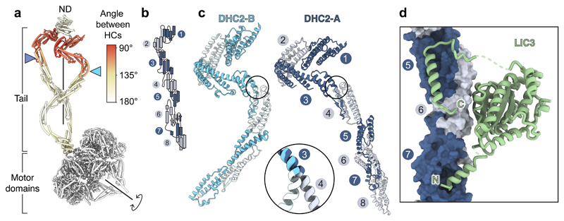
Figure 2. DHC2 asymmetry and LIC3 binding.
(a) Cylinder representation of the two copies of the DHC2, coloured by the rotation angle relating them. Other subunits omitted for clarity. Blue arrowheads indicate how equivalent helical bundles in DHC2TAIL are offset along the rotation axis. ND; N-terminal domain. (b) Topology of helical bundles (numbered) in the tail region. (c) Different conformations of the two copies of DHC2, which are shown after alignment on bundle 3. Enlargement shows major hinge site between bundles 3 and 4. (d) LIC3 binds to bundles 5-7 of the DHC2.

