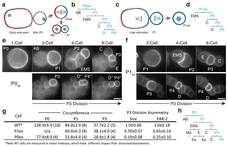
Figure 6. Premature loss of polarity and division asymmetry in P lineage cells derived from cell fragments.
(a) Laser-mediated extrusion of a posterior fragment from early establishment phase embryos containing both centrosomes yields a mini-P0 cell (P0ex) that undergoes normal asymmetric P0-like division to give rise to an AB:P1 cell pair. (b) Lineage derived from P0ex. Division pattern is normal until P3 (see h for wild type), which undergoes a symmetric division to yield two symmetric daughters, denoted D*/P4*. Blue indicates inheritance of the P lineage marker PAR-2. See stills in (e). (c) Extrusion of a posterior fragment during P0 cytokinesis instead yields a P1-like cell (P1ex). (d) Lineage derived from P1ex. Division pattern is normal through division of P3, which undergoes an asymmetric division as in wild type. See stills in (f). (e) An extruded mini P0 cell undergoes normal asymmetric divisions through birth of P3, which then divides symmetrically. Stills show 1-, 2-, 4-, and 8-cell equivalent stages, followed by the symmetric division of P3. The resulting daughters (P4* and D*) are labeled according to their position relative to C and E descendants, but denoted by * to indicate symmetric division. (f) An extruded P1 cell (P1ex) exhibits normal asymmetric divisions, including asymmetric division of P3. Stills show P1 and its descendants at the equivalent of the 2-, 4-, and 8-cell stages, followed by polarisation and asymmetric division of P3. Cell fragments in (e) and (f) were obtained from adjacent embryos mounted together on the same coverslip. Further examples in Supplementary Figure S5. Scale bars, 10 µm. For (e-f), see also Movie S4. (g) Table of extruded cell sizes and division asymmetries. Sample size indicated in parentheses. Mean ± STD shown. (h) Wild-type cell lineage showing division pattern of the 1- to 16-cell stage with cell identities indicated.

