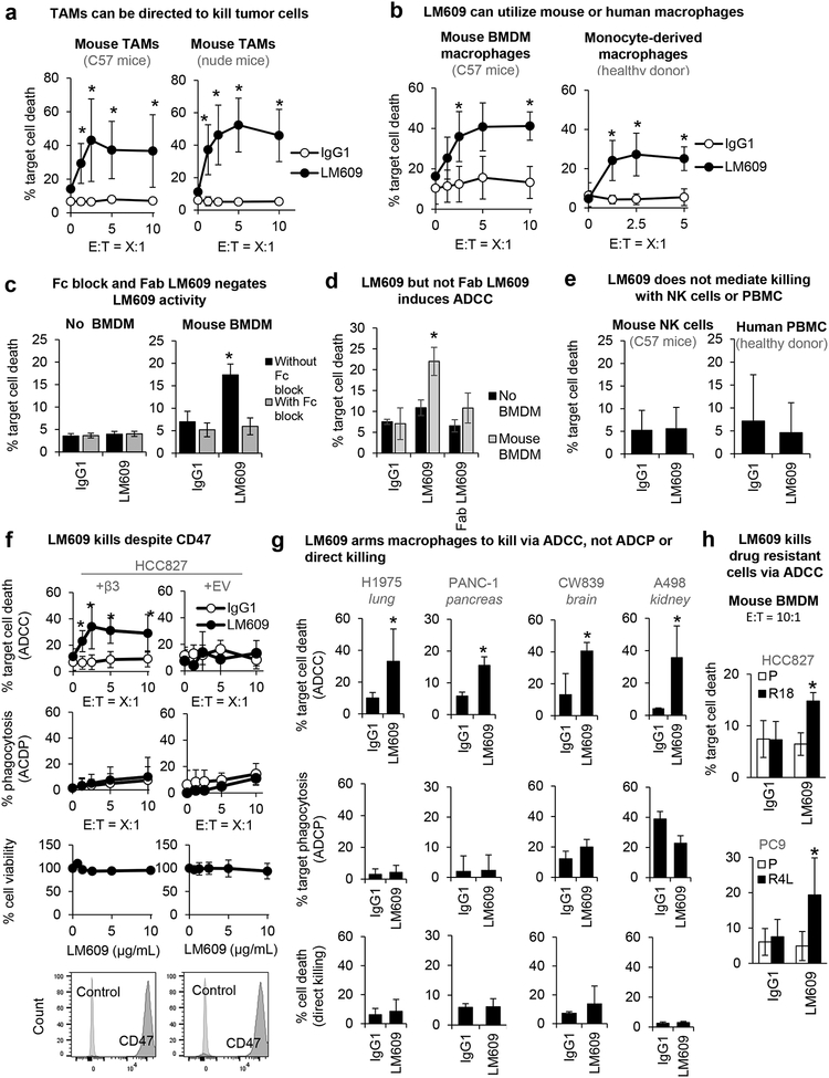
Figure 3: LM609 induces macrophage-mediated ADCC, not phagocytosis.
(A) Mouse tumor-associated macrophages (TAMs) are tested for ADCC activity. Graphs show mean±SD of % target cell (HCC827+β3) death at indicated effector-to-target cell ratios (E:T) induced by anti-αvβ3 LM609 (10 μg/mL) vs. IgG control. *P<0.05 compared to IgG1 using Student’s t test (repeated at least 3 times). Error bars indicate standard deviation.
(B) Human monocyte-derived macrophages (donor 631) and mouse bone marrow-derived macrophages (BMDM) are tested for their effects on target cells (HCC827+β3) for a range of E:T ratios. Tumor cell killing induced by anti-αvβ3 LM609 (10 μg/mL) vs. IgG control was measured. Error bars indicate standard deviation. *P<0.05 compared to the IgG1 group using Student’s t test (repeated at least 3 times).
(C) Death of target cells (HCC827+β3) induced by LM609 (10 μg/mL) in the absence or presence of BMDM (E:T=10:1) was evaluated in the absence or presence of function blocking antibodies targeting Fcγ receptors (Fc block). Error bars indicate standard deviation. *P<0.05 compared to IgG using Student’s t test (n=4).
(D) Death of target cells (HCC827+β3) induced by LM609 or Fab LM609 (10 μg/mL) in the absence or presence of BMDM (E:T=10:1) was evaluated. Error bars indicate standard deviation. *P<0.05 compared to IgG using Student’s t test (n=4).
(E) Death of target cells (HCC827+β3) measured after the co-incubation with mouse NK cells (E:T=5:1) and human PBMCs (E:T=500:1) with and without LM609 (10 μg/mL). Error bars indicate standard deviation.
(F) ADCC, ADCP, MTT assays, and flow cytometry for CD47 expression were performed for HCC827 cells with ectopic expression of empty vector (+EV) or the integrin β3 subunit (+β3). Death of target cells induced by LM609 (10 μg/mL) in the presence of mouse BMDM was evaluated at a range of E:T ratios (ADCC). Phagocytosis of target cells induced by LM609 (10 μg/mL) in the presence of mouse BMDM was evaluated (ADCP). Death of target cells induced by LM609 without effector cells was evaluated (% cell viability). CD47 expression on the target cells are shown. Error bars indicate standard deviation. *P<0.05 compared to IgG using Student’s t test (n= at least 3).
(G) ADCC, ADCP, and viability assays were performed for a panel of cancer cell lines with endogenous αvβ3 expression. Death of target cells induced by LM609 (10 μg/mL) in the presence of mouse BMDM was evaluated at an E:T ratio of 5:1 (ADCC). Phagocytosis of target cells induced by LM609 (10 μg/mL) in the presence of mouse BMDM was evaluated (ADCP). Direct killing of target cells by LM609 (10 μg/mL) was evaluated by measuring PI-positive cells after the 5 hr incubation of the target cells and LM609. Error bars indicate standard deviation. *P<0.05 compared to IgG using Student’s t test (n= at least 3).
(H) ADCC assay was performed for drug resistant cells with endogenous αvβ3 expression and drug sensitive cells with no αvβ3. Death of target cells induced by LM609 (10 μg/mL) in the presence of mouse BMDM was evaluated at an E:T ratio of 5:1. *P<0.05 compared to IgG using Student’s t test (n= at least 3).

