
Figure 3. Functional consequence of adaptor protein reorganization.
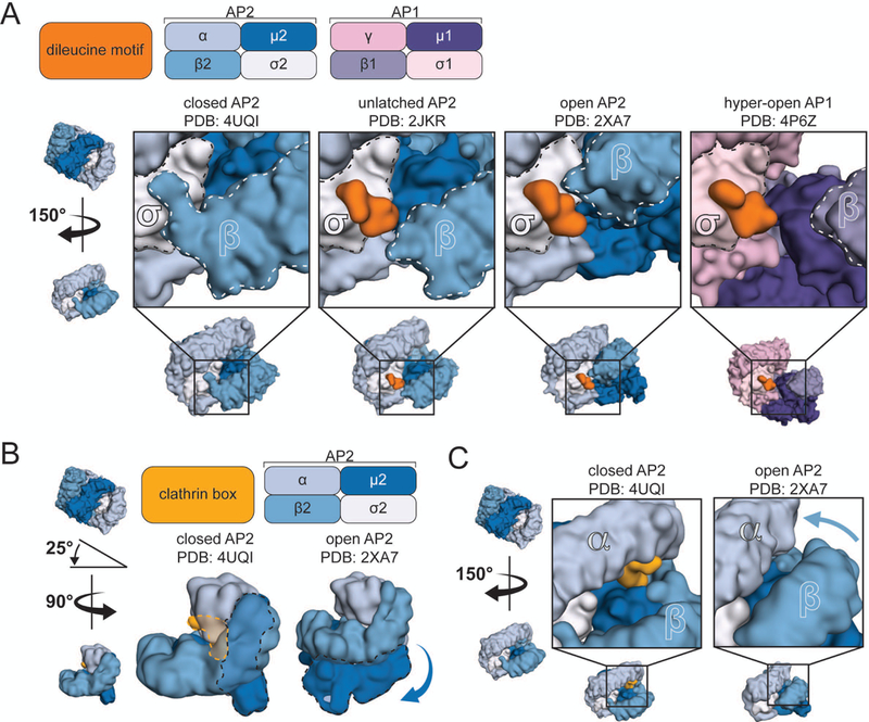
A. Close-up view of the dileucine motif binding pocket in four different conformations (indicated above). B and C. Interaction of the clathrin-binding box with the closed AP2 core. Positions of the μ2 subunit and clathrin-binding box within two configurations of the core (B, dashed lines). In the open conformation, the μ2 subunit has pivoted around the core (B, arrow) and the β2 solenoid has approached that of α (C, arrow), potentially excluding the clathrin-binding box.