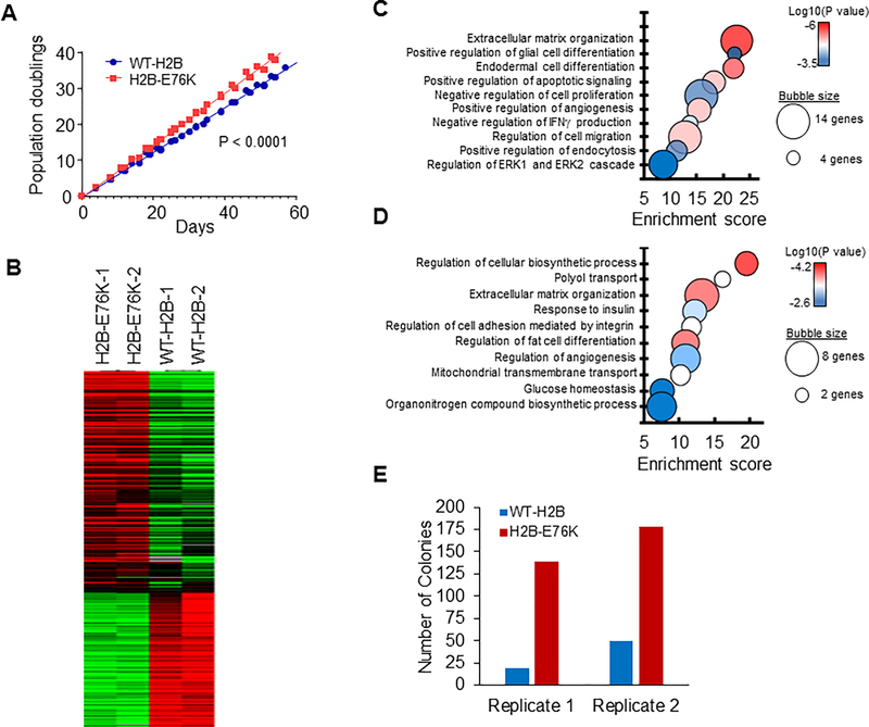
Figure 5. Expression of H2B-E76K alters growth properties and gene expression in human mammary epithelial cells.
(A) Proliferation of MCF10A transduced with lentivirus expressing either WT or H2B-E76K was measured every 3 to 4 days. The results from three different infections are shown. Linear regression analysis was used to calculate statistical significance. (B) Heat maps depicting differential gene expression between MCF10A cells expressing either WT or H2B-E76K. (C and D) Bubble plots of gene ontology analysis identify biological processes significantly different in cells expressing H2B-E76K compared to cells expressing WT H2B. Bubble area is relative to the number of genes identified in each classification. Ontology of genes with increased (C) or decreased (D) expression in cells expressing H2B-E76K. (E) MCF10A cells expressing WT H2B or H2B-E76K and PIK3CA(H1047R) were grown in soft agar for 14 days and colonies counted.

