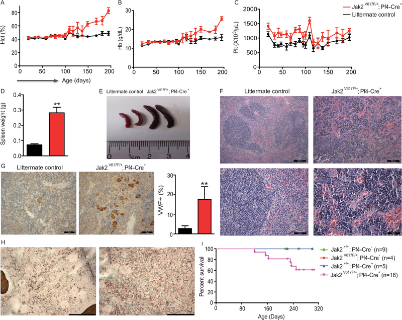
Figure 2.
Mk-specific expression of Jak2V617F leads to polycythemia. (A-C) Peripheral blood counts of Jak2 V617F/+; Pf4-Cre+ mice and littermate controls were monitored for 200 days. Jak2 V617F/+; Pf4-Cre+ mice showed significant increases in (A) hematocrit, (B) hemoglobin, and (C) platelet counts over the monitoring period (P < 0.01, by two-way ANOVA). (D-E) Jak2V617F/+; Pf4-Cre+ mice also displayed splenomegaly (increased spleen weight (D) and size (E)). (F) H&E staining revealed histopathological changes in splenic architecture (Scale bars depict 100 microns in the upper panel, 50 microns in the lower panel), as well as (G) increased VWF+ cells (Scale bars = 50 microns). (H) Reticulin staining revealed bone marrow fibrosis (Scale bars = 50 microns). In (D-H), mice were female and 6 months old. Results are representative of 2 independent experiments. N=6 animals per group. Bar graphs and line graphs depict mean ± SEM. (I) Kaplan-Meier survival curve (p=0.0374).

