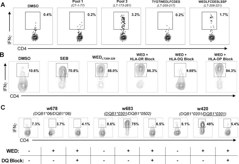
Figure 2: The WEDLT209–228 epitope is presented by HLA-DRB1*0401, HLA-DRB1*0301, and HLA-DQB1*0301 alleles.
A: ICS screening of expanded TILs from an MCC patient biopsy incubated with four MCPyV T-antigen 13-mer peptide pools (two shown). Reactive pool 3 was broken down into individual 13-mer peptides (two shown). The percentage of viable CD4+IFNγ+ T cells (negative for CD8/CD14/CD19) in the lymphocyte forward/side scatter region are denoted. Negative control: TILs stimulated with DMSO and the peptide ‘TYGWEDLFCDES’, which is 4 amino acids N-terminal to the ‘WED” peptide. Positive control: TILs stimulated with SEB. B: A WEDLT209–228-specific clone ± anti-HLA-class II locus-specific blocking monoclonal antibodies established HLA-DQ as the restricting locus. The percentage of viable CD4+IFNγ+ T cells (negative for CD8/CD14/CD19) in the lymphocyte forward/side scatter region are denoted. Negative control: DMSO-stimulated; Positive control: SEB-stimulated. C: HLA-DQB1*0301 restriction was established using LCLs from three different patients (“w678”, “w683,” and “w420”) with defined HLA-DQ genotype with a WEDLT209–228-specific CD4+ T-cell clone, peptide (“WED”), and inclusion of anti–HLA-DQ mAb (“DQ-Block”). The percent of viable CD4+IFNγ+ T cells (negative for CD8/CD14/CD19) in the lymphocyte forward/side scatter region are denoted. DMSO and SEB were used as negative and positive controls, respectively. Plots shown represent one of two replicates.

