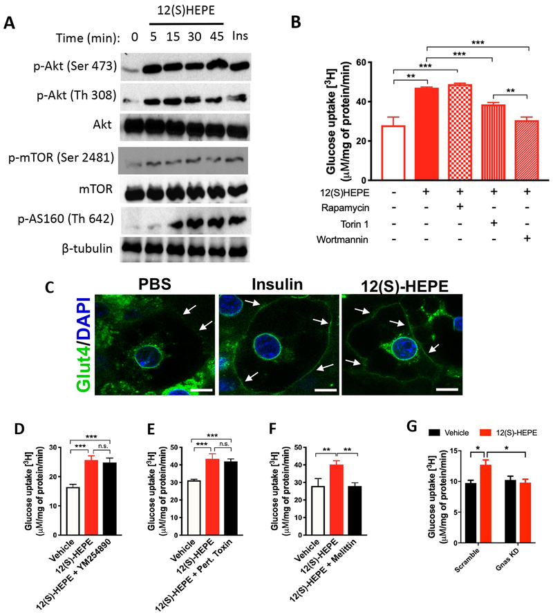
Figure 7: 12-HEPE promotes glucose uptake via a GsPCR/PI3K/AKT axis.
A) Western blotting for phospho-AKT (Ser473 and Th308), Akt, phospho-mTORC2 (Ser2481), mTOR, phospho-AS160 (Th642), and β-tubulin (loading control) in mouse brown adipocytes treated with 12(S)-HEPE or vehicle.
B) In vitro glucose uptake into murine brown adipocytes treated with 12(S)-HEPE or vehicle in the presence or absence of pre-treatment with rapamycin, Torin 1 or Wortmannin.
C) Immunofluorescent staining for Glut-4 in murine brown adipocytes treated with PBS, insulin or 12(S)-HEPE. The white arrows indicate Glut-4 staining in the plasma membrane.
D-F) In vitro glucose uptake into murine brown adipocytes treated with 12(S)-HEPE or vehicle in the presence or absence of pre-treatment with YM254890, Pertussis Toxin or Melittin.
G) In vitro glucose uptake into Scramble (control) or Gnas KD murine brown adipocytes treated with 12(S)-HEPE or vehicle. *P<0.05, **P<0.01, ***P<0.001. Data are represented as mean ± S.E.M. See also Figure S7.

